What is femoral acetabular impingement (FAI)?
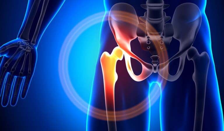
Femoral acetabular impingement (or Femoroacetabular Impingement) is a condition of the hip in which there is abnormal contact between the femoral head and the acetabulum (hip socket). This abnormal contact can lead to hip labral tears and damage to the cartilage surfaces of the hip.

Photo from orthoinfo.aaos.org
What are the types of FAI?
CAM impingement, pincer impingement, and mixed impingement. The bony abnormalities that occur that lead to FAI are common in the general population and are often asymptomatic. They typically develop throughout normal childhood growth and development, but there are genetic components, as well as certain conditions or injuries that can lead to the bony overgrowth and impingement.
CAM impingement refers to femoral based disorders where there is an excess of bone at the head/neck junction of the femur leading to impingement on the acetabular rim during hip flexion.

Pincer impingement refers to acetabular based disorders where there can be increased bony overgrowth on the acetabular rim, or increased overhang of normal bone due to abnormal positioning of the hip socket.

Mixed impingement refers to combination of both CAM and pincer lesions and is present in up to 80% of cases.

CAM, PINCER, MIXED photos from williamjbose.com
What can I expect from FAI?
The altered bony morphology ultimately leads to altered hip biomechanics. When the bone impinges, it pinches the hip labrum and can damage it. The labrum is a ring of cartilage tissue that attaches to the acetabular ring and acts like a gasket to provide more stability to the hip joint, as well as to create a “suction-seal” of the joint to allow for smoother, more fluid motion of the joint. When this tissue is damaged and the suction-seal is broken, this can lead to further damage to the hip joint cartilage and cause pain. Ultimately, the natural history of FAI leads to early onset hip dysfunction and arthritis.
What are the symptoms of FAI?
Symptoms of FAI and hip labral tears often present as groin pain, exacerbated by hip flexion. Patients sometimes report pain with sitting and pain into the lateral hip and buttock, and often with labral tears will complain of mechanical symptoms of clicking or popping. Symptoms can develop specifically after injuries, but can occur more insidiously over time without injury. New onset symptoms however do not mean that the FAI just began as the bony abnormalities were likely present for years. The development of symptoms is likely attributable to a progression of injury, or an acute exacerbation by a new injury.
How is FAI diagnosed?
Diagnosis is achieved with a good history of symptoms and thorough physical exam. Xrays are a big part of the diagnosis and confirm the presence of bony problems such as CAM or pincer lesions. MRI is used to better evaluate for labral tearing or cartilage injury and sometimes CT scans with 3D reconstructions are necessary for pre-surgical planning.

Xray showing CAM and pincer

MRI showing labral tear – red arrow

3D CT Scan showing femoral CAM lesion
Femoral Acetabular Impingement Treatment
Treatment of FAI always begins with conservative treatment including rest, anti-inflammatory medications, and physical therapy program. The goal of therapy is not to improve hip motion or change the bony morphology, but instead to increase strength of the core, gluteal muscles and hip flexors to decrease the joint reactive forces on the hip and thus decrease pain and symptoms. Injections can also be used to help with symptoms, and as a diagnostic tool to confirm that the pain is coming from the hip joint. When these conservative treatments fail, surgery may be indicated.
What to expect from FAI surgery
Surgery for FAI involves hip arthroscopy where the hip joint is accessed through tiny incisions and a camera. The hip labrum can be repaired as well as the excess bone from CAM and pincer lesions resected to improve hip motion, remove impingement, and re-store the “suction-seal” of the hip joint. In some cases of severe labral damage, the hip labrum needs to be reconstructed with use of a tendon graft. When done in the appropriate setting, this surgery is very successful in alleviating symptoms and in helping to preserve the hip joint from further cartilage damage. In some cases, if the FAI has not been caught early enough, the cartilage damage (arthritis) can be too severe. In these cases, hip replacement may be the only surgical option if other treatments fail, however, this is typically reserved for older patients, ideally over 60 if possible.

A – Labral tear. B – Labral repair. C- labral repair with restoration of “suction-seal”. D – labral repair
What is the average recovery time?
Recovery from hip arthroscopy typically takes 4-6 months. This usually requires a period of time on crutches with limitation of activities. Physical therapy post surgery is a must to regain strength, proper motion, and full function for optimal outcomes.
Surgical treatment of FAI with hip arthroscopy is a complex procedure which requires specialty training. At OMG, Drs. Sando and Watson are well versed in treating this pathology and with the latest techniques of complex hip arthroscopy. Come visit one of them for a consultation regarding your hip pain!
Dr. Mark Sando is an internationally recognized orthopaedic surgeon specializing in sports medicine, arthroscopy and injuries of the shoulder, knee and hip. He earned his medical degree from Case Western Reserve University School of Medicine and completed residency at the University of Maryland Medical Center and R Adams Cowley Shock Trauma Center. After residency, Dr. Sando went on to complete subspecialized training in Sports Medicine as a fellow at the prestigious Kerlan-Jobe Orthopaedic Clinic in Los Angeles, CA under the direction of Dr. Neal ElAttrache. He has worked with numerous college athletic programs and professional teams including the world champion Los Angeles Kings, Los Angeles Lakers, Los Angeles Dodgers, and Los Angeles Sparks. Dr. Sando has been with the Orthopaedic Medical Group of Tampa Bay since 2015. Full Bio